Medical Science
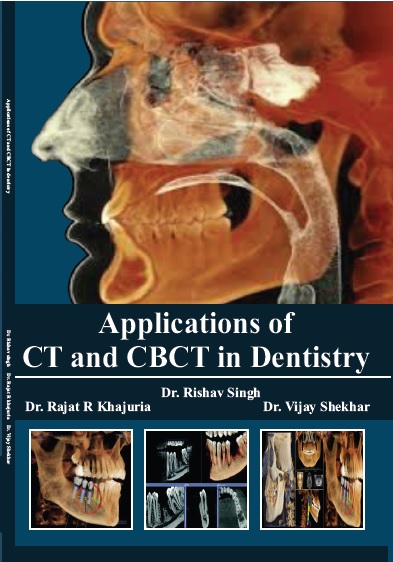
Applications Of Ct And Cbct In Dentistry.
by Dr. Rishav Singh
ISBN Number : 978 - 1- 73030 - 082 - 0
Authors Details
| Author Name | Image | About Author |
|---|---|---|
| Dr. Rishav Singh |  |
Tutor, Pediatric and Preventive Dentistry, Dental Institute, Rajendra Institute
of Medical Sciences, Ranchi, Jharkhand. |
| Dr. Vijay Shekhar |  |
Reader, Conservative Dentistry and Endodontics, Hazaribag College of Dental Sciences and hospital, Jharkhand. |
| Dr. Rajat R Khajuria |  |
Lecturer, Prosthodontics including Crown and Bridge,
Indira Gandhi Govt. Dental College, Jammu |
Book Description
Computed tomography (CT), originally known as computed axial tomography (CAT or CT scan) and body section roentgenography, is a medical imaging method employing tomography where digital geometry processing is used to generate a three dimensional image of an object from a large series of two dimensional x-ray images taken around a single axis of rotation. The word “tomography” is derived from the Greek “tomos” (slice) and “graphein” (to write). CT produces a volume of which can be manipulated, through a process known as windowing, in order to demonstrate various structure based on the their ability to attenuate x-ray beam. It is a medical imaging procedure that utilizes computer-processed X-rays to produce tomographic images or 'slices' of specific areas of the body. These cross-sectional images are used for diagnostic and therapeutic purposes in various medical disciplines. Digital geometry processing is used to generate a three-dimensional image of the inside of an object from a large series of two-dimensional X-ray images taken around a single axis of rotation. CT produces a volume of data that can be manipulated, through a process known as "windowing", in order to demonstrate various bodily structures based on their ability to block the X-ray beam. Although historically the images generated were in the axial or transverse plane, perpendicular to the long axis of the body, modern scanners allow this volume of data to be reformatted in various planes or even as volumetric (3D) representations of structures. Although most common in medicine, CT is also used in other fields, such as nondestructive materials testing. Another example is archaeological uses such as imaging the contents of sarcophagi. Until recently, dentists have evaluated the jaw predominantly by using radiographs in their office. The development of dental computed tomographic (CT) reformatting programs, however, has completely revolutionized and changed the fashion in which we radiographically evaluate the jaw today. Today, these programs are used to evaluate patients with dental implants; in addition, they are being used to assess tumors , cysts , inflammatory disease , oroantral fistulas , silicone implants , fractures , and surgical procedures. The programs are useful because they provide accurate information about the height and width of the jaw, as well as information about the location of vital structures, such as the mandibular canal, mental foramen, mandibular foramen, incisive foramen, and maxillary sinuses. In addition, detailed information about internal anatomy and the relationship between lesions and the cortical margins and roots of the teeth can be established. These programs are optimally used as an adjunct to, rather than a substitute for, conventional dental radiography. X Ray Computed Tomography (CT) has experienced tremendous growth in recent years, in terms of both basic technology and new clinical applications. Much advancements has been achieved in major CT components such as tube, detector, slip ring, data acquisition systems and algorithms. Many new clinical applications have been developed since the introduction of helical and multislice CT.


